बोटोनिएरे विकृति क्या है?
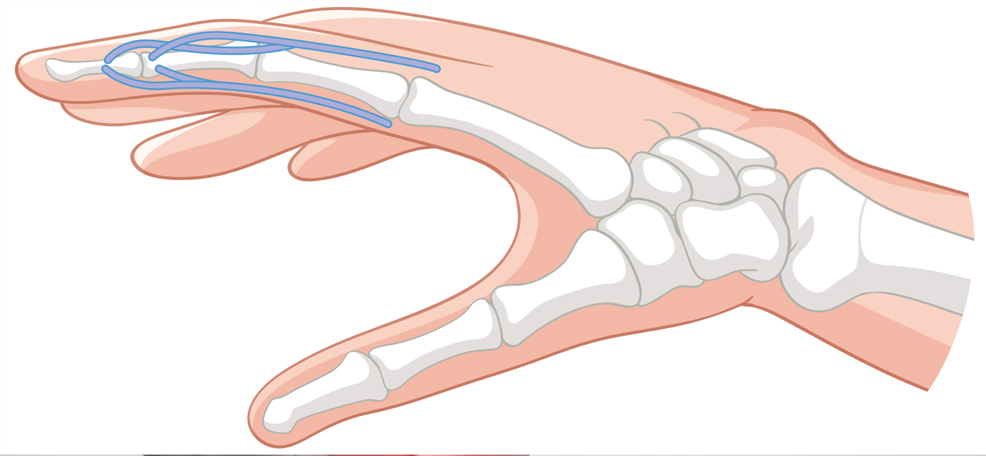
Boutonniere विकृति, जिसे बटनहोल विकृति या केंद्रीय पर्ची चोट के रूप में भी जाना जाता है, उंगलियों की स्थिति है — शायद ही कभी पैर की उंगलियों — जिसमें प्रोक्सिमल इंटरफालैंगियल (पीआईपी) संयुक्त एक फ्लेक्स (बेंट) स्थिति में आयोजित किया जाता है जबकि डिस्टल इंटरफालैंगियल (डीआईपी) संयुक्त अतिविस्तारित (बेंट बैकवर्ड) हो जाता है। परिणाम प्रभावित उंगली की एक विशेषता crooked उपस्थिति और पूरी तरह से इसे सीधा करने में असमर्थता है।
'boutonnière' शब्द 'buttonhole' के लिए फ्रेंच है, जो तब उत्पन्न होने वाली उपस्थिति का जिक्र करता है जब बाहरी सेंसर टेंडन टियर्स की केंद्रीय पर्ची और पार्श्व बैंड के बीच की खाई के माध्यम से पीआईपी संयुक्त protrudes — एक बटनहोल के माध्यम से एक बटन जैसा दिखता है।
विकृति तब विकसित होती है जब एक्स्टेंसर टेंडन की केंद्रीय पर्ची — जो पीआईपी संयुक्त को सीधा करने के लिए जिम्मेदार है — बाधित है। यह व्यवधान पार्श्व बैंड को अस्थिर रूप से स्थानांतरित करने का कारण बनता है, जिसके परिणामस्वरूप मजबूर पीआईपी flexion और डीआईपी अति-विस्तार होता है। यदि 3 सप्ताह से अधिक के लिए अनुपचारित छोड़ दिया जाता है, तो विकृति निश्चित हो सकती है और इसे सही करने में काफी कठिनाई होती है।
बोटोनिएरे विकृति के कारण
बौटोनिएर विकृति विभिन्न प्रकार के दर्दनाक, भड़काऊ और जन्मजात स्थितियों से उत्पन्न हो सकती है। सबसे आम कारणों में शामिल हैं:
- दर्दनाक चोट: मध्य उंगली के संयुक्त, एक लेसरेशन, क्रश चोट, या पीआईपी संयुक्त के विघटन की डोर्सल (बैक) सतह पर एक बलपूर्वक झटका, केंद्रीय पर्ची के थंडन को तोड़ सकता है या निकाल सकता है। यह सबसे लगातार कारण है।
- Rheumatoid गठिया (RA): पीआईपी संयुक्त में पुरानी सूजन प्रगतिशील कमजोरी और केंद्रीय पर्ची के घटना टूटने की ओर जाता है। Synovitis fibrous ऊतक buildup, बोनी कटाव, और उपास्थि क्षति का कारण बनता है, धीरे-धीरे जोड़ों को विकृत करता है।
- ऑस्टियोआर्थराइटिस: उंगली के जोड़ों में अपक्षयी परिवर्तन बाहरी तंत्र को कमजोर कर सकते हैं और समय के साथ विकृति में योगदान कर सकते हैं।
- बर्न्स: उंगली के dorsum के लिए थर्मल चोटों का कारण बन सकता है ठेकेदारों और tendon क्षति के कारण Boutonniere विकृति होती है। तनाव इस्केमिया जलने की चोटों में तनाव टूटने के लिए एक संभावित तंत्र है।
- फिंगर फ्रैक्चर: मध्य phalanx को शामिल करने वाले फ्रैक्चर, विशेष रूप से केंद्रीय स्लिप अटैचमेंट के एवल्शन फ्रैक्चर, इस विकृति का उत्पादन कर सकते हैं।
- खेल चोट: संपर्क खेल में जमी हुई उंगलियों जैसे कि बास्केटबॉल, फुटबॉल और वॉलीबॉल में अक्सर केंद्रीय स्लिप चोटों को शामिल किया जाता है जो बिना मान्यता प्राप्त होने पर बाउटोनियर विकृति में विकसित हो सकता है।
- पीआईपी संयुक्त विघटन: एक ज्वालामुखी पीआईपी विच्छेदन मध्यम phalanx के डॉर्सल होंठ को नष्ट कर सकता है, जो केंद्रीय पर्ची लगाव को बाधित कर सकता है।
जन्मजात कारक: दुर्लभ मामलों में, जन्मजात एप्लासिया या उंगली के एक्स्टेंसर के हाइपोप्लासिया के परिणामस्वरूप जन्म से बौथी विकृति हो सकती है।
बोटोनीरे विकृति के लक्षण और लक्षण
लक्षण तुरंत एक तीव्र चोट के बाद विकसित हो सकते हैं या 1 से 3 सप्ताह में धीरे-धीरे उभर सकते हैं क्योंकि पार्श्व बैंड प्रगतिशील रूप से माइग्रेट हो सकते हैं। विशेषता विशेषताओं में शामिल हैं:
- पीआईपी संयुक्त फ्लेक्सियन विकृति: प्रभावित उंगली का मध्य संयुक्त एक मुड़ने की स्थिति में फंस गया है और सक्रिय रूप से सीधा नहीं किया जा सकता है — Boutonniere विकृति का संकेत।
- डीआईपी संयुक्त उच्च रक्तचाप: पार्श्व बैंड के विस्थापन के कारण उंगलियों को असामान्य रूप से पिछड़े (hyperextends) झुकता है और टर्नऑन मैकेनिक्स को बदल देता है।
- दर्द और कोमलता: दर्द आम तौर पर पीआईपी संयुक्त की डॉर्सल सतह पर महसूस किया जाता है, जिससे उंगली को बढ़ाने के प्रयासों से बदतर हो जाता है।
- सूजन और एडेमा: उंगली के मध्य जोड़ों पर स्थानीयकृत सूजन, विशेष रूप से चोट के बाद तीव्र चरण में।
- पकड़ शक्ति का नुकसान: बदली हुई उंगली यांत्रिकी और दर्द के कारण कठिनाई को पकड़ना या pinching ऑब्जेक्ट।
- कठोरता: पीआईपी संयुक्त की प्रगतिशील कठोरता, विशेष रूप से पुरानी या अनुपचारित मामलों में।
- कार्यात्मक सीमा: लेखन, बटनिंग कपड़े, या टाइपिंग जैसे ठीक मोटर कार्यों को करने में कठिनाई।
पैथोलॉजी
Boutonniere विकृति का पैथोलॉजिकल आधार जोन III (PIP संयुक्त के स्तर पर) पर बाहरी तंत्र के विघटन में निहित है। जब केंद्रीय पर्ची टूट जाती है या तबाह हो जाती है, पार्श्व बैंड अपने डोर्सल स्थिरीकरण को खो देते हैं और पीआईपी संयुक्त के घूर्णन की धुरी के नीचे गुजरते हैं। पीआईपी संयुक्त के विस्तार के बजाय, वे अब फ्लेक्सर्स के रूप में कार्य करते हैं। इसके साथ ही, वे डीआईपी संयुक्त पर अपने पुल को पेश करना जारी रखते हैं, जो अति-विस्तार का उत्पादन करते हैं। Reumatoid गठिया में, synovitis उत्तरोत्तर erodes periarticular नरम ऊतक, जब तक यह सामान्य कार्यात्मक भार के तहत टूट जाता है केंद्रीय पर्ची कमजोर।
बोटोनीरे विकृति का निदान
शीघ्र और सटीक निदान महत्वपूर्ण है, क्योंकि 3 सप्ताह से अधिक देरी से उपचार काफी जटिल है। निम्नलिखित नैदानिक दृष्टिकोण का उपयोग किया जाता है:
नैदानिक परीक्षा
एक गहन इतिहास और शारीरिक परीक्षा प्राथमिक नैदानिक उपकरण है। चिकित्सक आराम से उंगली की मुद्रा का मूल्यांकन करता है, पीआईपी और डीआईपी जोड़ों की गति की सक्रिय और निष्क्रिय सीमा, और केंद्रीय पर्ची सम्मिलन पर कोमलता।
Elson Test
केंद्रीय पर्ची अखंडता के लिए एक विशिष्ट नैदानिक परीक्षण। पीआईपी संयुक्त एक तालिका के किनारे पर फ्लेक्सियन के 90 डिग्री पर आयोजित किया जाता है और रोगी को प्रतिरोध के खिलाफ उंगली का विस्तार करने के लिए कहा जाता है। यदि डीआईपी संयुक्त लचीला और फ्लॉपी रहता है, तो केंद्रीय पर्ची अखंडता बरकरार है। यदि इस युद्ध के दौरान डीआईपी संयुक्त कठोर हो जाता है, तो केंद्रीय पर्ची विघटन की पुष्टि की जाती है। यह प्रारंभिक निदान के लिए सबसे विश्वसनीय बेडसाइड परीक्षण है।
एक्स-रे (रेडियोग्राफ)
Anteroposterior और पार्श्व रेडियोग्राफ का उपयोग मध्य phalanx के डोर्सल बेस पर केंद्रीय स्लिप सम्मिलन पर संबद्ध एवल्शन फ्रैक्चर की पहचान करने के लिए किया जाता है, किसी भी बॉनी विखंड विस्थापन का पता लगाता है, और पार्श्व दृश्यों पर डीआईपी संयुक्त अतिविस्तार की डिग्री का आकलन करता है।
MRI (Magnetic Resonance Imaging)
MRI विस्तृत सॉफ्ट टिशू इमेजिंग प्रदान करता है और नैदानिक परीक्षा असंगत होने पर उपयोगी होता है। यह केंद्रीय पर्ची और आसपास की संरचनाओं की अखंडता को सीधे देख सकता है।
अल्ट्रासाउंड
गतिशील अल्ट्रासाउंड आकलन आंदोलन के दौरान वास्तविक समय में एक्स्टेंसर टेंडन की कल्पना कर सकता है और केंद्रीय पर्ची विघटन की पहचान कर सकता है, विशेष रूप से तीव्र नैदानिक सेटिंग में उपयोगी है।
Boutonniere विकृति के लिए उपचार
उपचार विकृति की गंभीरता, क्रूरता और अंतर्निहित कारण पर निर्भर करता है। प्राथमिक लक्ष्य पीआईपी संयुक्त और सामान्य डीआईपी संयुक्त संरेखण के पूर्ण सक्रिय विस्तार को बहाल करना है। रूढ़िवादी प्रबंधन को प्राथमिकता दी जाती है और शुरू में शुरू होने पर सबसे प्रभावी होता है।
रूढ़िवादी (गैर शल्य चिकित्सा) उपचार
- छिड़काव: पीआईपी संयुक्त एक स्थिर स्प्लिंट (जैसे ओवल-8 या बंनेल सुरक्षा-पिन स्प्लिंट) का उपयोग करके पूर्ण विस्तार में immobilized है जबकि डीआईपी संयुक्त फ्लेक्स के लिए स्वतंत्र है। तीव्र चोटों के लिए 6-8 सप्ताह के लिए स्प्लिन्टिंग लगातार बनाए रखा जाता है, और पुराने मामलों के लिए 12 सप्ताह तक। इस अवधि के दौरान गलती से फ्लेक्स करने के लिए उंगली की अनुमति सभी प्रगति को उलट सकती है।
- डीआईपी संयुक्त व्यायाम: जबकि पीआईपी संयुक्त को विस्तार में विभाजित किया जाता है, डीआईपी संयुक्त सक्रिय रूप से फ्लेक्स किया जाता है और पूरे दिन विस्तारित होता है। ये अभ्यास पार्श्व बैंड को निष्क्रिय रूप से अपनी सही स्थिति में वापस लाने में मदद करते हैं।
- दवा: दर्द और सूजन प्रबंधन के लिए NSAIDs और एनाल्जेसिक। संधिशोथ से संबंधित विकृति में, रोग-संशोधित एंटीरहेमेटिक ड्रग्स (DMARD) और कॉर्टिकोस्टेरॉइड इंजेक्शन प्रणालीगत सूजन को नियंत्रित करने के लिए इस्तेमाल किया जा सकता है।
- रात्रि: प्राथमिक इमोबिलाइजेशन अवधि के बाद, जारी रखा गया है कि पीआईपी विस्तार लाभ को बनाए रखने के लिए कई अतिरिक्त सप्ताहों के लिए रात की स्प्लिनिंग की सिफारिश की जा सकती है।
शल्य चिकित्सा उपचार
जब रूढ़िवादी उपचार विफल हो जाता है, तो सर्जरी का संकेत दिया जाता है, जब एक गंभीर चालन होता है, एक बड़ा विस्थापित बोनी टुकड़ा, या जब विकृति संयुक्त विनाश के साथ संधिशोथ के कारण होती है। सर्जिकल विकल्पों में शामिल हैं:
- केंद्रीय पर्ची मरम्मत या पुनर्निर्माण: ruptured केंद्रीय पर्ची शल्य चिकित्सा रूप से प्राथमिक सिवनी या टेंडन grafting का उपयोग कर मरम्मत की है। एक avulsed bony टुकड़ा एक हड्डी एंकर या पिन निर्धारण का उपयोग करके फिर से संलग्न है।
- पीआईपी संयुक्त पिनिंग: एक अस्थायी किर्स्चनर तार (K-wire) को पीआईपी संयुक्त में डाला जाता है ताकि इसे 4-6 सप्ताह तक पूर्ण विस्तार में बनाए रखा जा सके।
- एक्स्टेंसर टेनोटॉमी: क्रोनिक Boutonniere विकृति में, डीआईपी संयुक्त में टर्मिनल एक्स्टेंसर थंडन का एक टेनोटॉमी हाइपरएक्सटेंशन को कम कर सकता है और उंगली संतुलन में सुधार कर सकता है।
- संयुक्त प्रतिस्थापन (Arthroplasty): Boutonniere विकृति के लिए माध्यमिक से उन्नत संधिशोथ के लिए संयुक्त विनाश के साथ, सिलिकॉन पीआईपी संयुक्त प्रतिस्थापन संरेखण और कार्य को बहाल कर सकता है।
- संयुक्त संलयन (Arthrodesis): कार्यात्मक स्थिति में पीआईपी संयुक्त का फ्यूजन दर्द को समाप्त करता है और संयुक्त गति के स्थायी नुकसान की लागत से प्रगतिशील विकृति को रोकता है। अंतिम चरण के मामलों के लिए आरक्षित।
सर्जरी के बाद, रोगी कई हफ्तों के लिए एक स्प्लिंट पहनते हैं और एक संरचित से गुजरते हैं भौतिक चिकित्सा गतिशीलता और ताकत को बहाल करने के लिए कार्यक्रम।
बोटोनिएर विकृति के लिए फिजियोथेरेपी उपचार
फिजियोथेरेपी दोनों रूढ़िवादी और पोस्ट-सर्जिकल प्रबंधन के लिए अनिवार्य है। एक व्यक्तिगत पुनर्वास कार्यक्रम दर्द, सूजन, संयुक्त गतिशीलता, ताकत और कार्यात्मक वसूली को संबोधित करता है।
इलेक्ट्रोफिजिकल एजेंट
- हीट थेरेपी: ऊतक को गर्म करने के लिए व्यायाम सत्र से पहले मोस्ट गर्मी या पैराफिन मोम स्नान लागू किया जाता है, हाथ की मांसपेशियों को आराम देता है, और टेंडन की व्यापकता में सुधार करता है।
- शीत थेरेपी / क्रायोथेरेपी: आइस पैक या कोल्ड कंप्रेस का उपयोग तीव्र चरण में किया जाता है ताकि पीआईपी संयुक्त के आसपास सूजन, दर्द और स्थानीय सूजन को कम किया जा सके।
- TENS (transcutaneous विद्युत तंत्रिका उत्तेजना): रीढ़ की हड्डी के स्तर पर तंत्रिका संकेतों को संशोधित करके दर्द को कम करता है, जिससे रोगी को व्यायाम चिकित्सा में आराम से भाग लेने की अनुमति मिलती है।
- अल्ट्रासाउंड थेरेपी: Therapeutic अल्ट्रासाउंड ऊतक चिकित्सा को बढ़ावा देता है, निशान ऊतक गठन को कम करता है, और उपासना और पुरानी चरणों में चालन extensibility में सुधार करता है।
- इंटरफेरेंशियल करंट थेरेपी (IFT): दर्द को कम करने, सूजन को कम करने और प्रभावित उंगली में स्थानीय परिसंचरण में सुधार के लिए गहरी पेनेट्रेटिंग विद्युत उत्तेजना प्रदान करता है।
- लो-लेवल लेजर थेरेपी (LLLT): सेलुलर मरम्मत को बढ़ावा देने के लिए इस्तेमाल किया जा सकता है और केंद्रीय पर्ची के आसपास पेरिटेनडिनस ऊतकों में सूजन को कम किया जा सकता है।
स्प्लिन्टिंग एंड ऑर्थोटिक मैनेजमेंट
फिजियोथेरेपिस्ट पूर्ण विस्तार में पीआईपी संयुक्त को बनाए रखने के लिए कस्टम थर्माप्लास्टिक splints बनाने और फिटिंग करने में एक केंद्रीय भूमिका निभाता है। स्प्लिन्टिंग प्रोटोकॉल को अंततः उपचार अग्रिमों के रूप में संशोधित किया जाता है — स्थैतिक से गतिशील स्प्लिंट तक संक्रमण जो उपचार की रोकथाम के लिए जारी रखते हुए गति की नियंत्रित रेंज की अनुमति देते हैं।
व्यायाम थेरेपी
- डीआईपी फ्लेक्सियन एक्सरसाइज (During Splinting): स्प्लिंट द्वारा विस्तार में आयोजित पीआईपी संयुक्त के साथ, रोगी सक्रिय रूप से डीआईपी गतिशीलता को बनाए रखने और पार्श्व बैंड के डोर्सल माइग्रेशन को बढ़ावा देने के लिए दिन भर डीआईपी संयुक्त को बार-बार बढ़ा देता है।
- पीआईपी एक्सटेंशन एक्सरसाइज: इमोबिलाइज़ेशन अवधि के बाद, सौम्य सक्रिय सहायता प्राप्त और सक्रिय पीआईपी एक्सटेंशन अभ्यास को एक्स्टेंसर टेंडन फंक्शन और गति की संयुक्त रेंज को बहाल करने के लिए पेश किया जाता है।
- टेंडन ग्लाइडिंग एक्सरसाइज: विशिष्ट हाथ की स्थिति (सीधे, हुक मुट्ठी, पूर्ण मुट्ठी, टेबलटॉप) के एक अनुक्रम ने फ्लेक्सर और एक्स्टेंसर टेंडन के स्वतंत्र ग्लाइडिंग को बढ़ावा देने और आसंजन गठन को रोकने के लिए दोहराए गए।
- व्यायाम को मजबूत करना: प्रोग्रेसिव रेसिस्टेंस एक्सरसाइज यूजिंग पोटीन, ग्रिप स्ट्रेंथ, चुटकी ताकत और आंतरिक मांसपेशी फंक्शन को बहाल करने के लिए गेंद को निचोड़ना।
- एक्सरसाइज: विस्तार में पीआईपी संयुक्त के सज्जन निष्क्रिय स्ट्रेचिंग और फ्लेक्सर और एक्स्टेंसर बलों के बीच संतुलन बहाल करने के लिए डीआईपी संयुक्त लचीलापन में।
मैनुअल थेरेपी
- संयुक्त मोबिलाइजेशन: संयुक्त कैप्सूल एक्स्टेंसिबिलिटी को बेहतर बनाने और गति की शारीरिक रेंज को बहाल करने के लिए पीआईपी और डीआईपी जोड़ों पर लागू कुशल निष्क्रिय जुटाने की तकनीक।
- शीतल ऊतक मालिश: मालिश और myofascial रिहाई तकनीक हाथ, thenar और hypothenar eminences की आंतरिक मांसपेशियों के लिए लागू किया, और dorsal tendon संरचनाओं तनाव को कम करने और रक्त प्रवाह में सुधार करने के लिए।
- निशान प्रबंधन: पोस्ट-सर्जिकल या पोस्ट-लेसरेशन के मामलों में, निशान मालिश, सिलिकॉन जेल शीटिंग और desensitization तकनीकों को आसंजन गठन को कम करने और निशान व्यवहार्यता में सुधार करने के लिए नियोजित किया जाता है।
कार्यात्मक पुनर्वास
As recovery progresses, functional retraining focuses on restoring the ability to perform activities of daily living (ADLs). Task-specific activities such as pinching, grasping, writing, and typing are progressively reintroduced to retrain coordinated finger function.
Occupational Therapy Integration
Occupational therapists work alongside physiotherapists to address ADL performance, adaptive equipment, workplace modifications, and vocational rehabilitation for patients whose daily activities or occupation have been impacted by the deformity.
रोगी शिक्षा
Education is an integral part of recovery from boutonniere deformity. Patients are counseled on the following:
- शर्त को समझना: Clear explanation of the anatomy of the extensor mechanism, the mechanism of injury, and why consistent splinting and exercises are essential to recovery.
- Importance of Early Treatment: Patients are informed that delaying treatment beyond 3 weeks significantly worsens outcomes and may make the deformity permanent or require more complex surgical intervention.
- Splint Compliance: Emphasis on wearing the splint continuously as prescribed — removing it to bathe or clean the splint — and never allowing the finger to flex at the PIP joint during the immobilization period, as even a single accidental bend can restart the healing clock.
- होम एक्सरसाइज प्रोग्राम: Instruction in a personalized set of daily exercises including DIP flexion, tendon gliding, and progressive strengthening to be performed independently between clinic sessions.
- गतिविधि संशोधन: Guidance on avoiding activities that risk re-injury to the finger during the healing phase. Return to sports is permitted only with appropriate protective splinting or buddy taping.
- Recognizing Warning Signs: Patients are educated on signs of deterioration — worsening deformity, increased pain, or numbness — that warrant prompt reassessment by their healthcare provider.
- Long-term Prognosis: Realistic discussion of recovery timelines (3–6 months for full recovery), the potential for residual stiffness, and the importance of ongoing hand exercises after formal rehabilitation ends.
संबंधित शर्तें
Boutonniere deformity shares clinical overlap with and may be associated with the following conditions:
- Rheumatoid गठिया: A major systemic cause of boutonniere deformity through progressive synovitis and central slip erosion.
- Swan Neck Deformity: The inverse finger deformity — PIP hyperextension and DIP flexion — also commonly seen in rheumatoid arthritis.
- Mallet Finger: Disruption of the terminal extensor tendon causing DIP flexion deformity; a related extensor tendon injury.
- Trigger Finger (Stenosing Tenosynovitis): Inflammation of the flexor tendon sheath causing locking or catching of the finger in flexion.
- Dupuytren’s Contracture: Progressive fibrosis of the palmar fascia causing fixed flexion deformity of the fingers.