What Is Boutonniere Deformity?
Boutonniere deformity, also known as buttonhole deformity or central slip injury, is a condition of the fingers — and rarely the toes — in which the proximal interphalangeal (PIP) joint is held in a flexed (bent) position while the distal interphalangeal (DIP) joint becomes hyperextended (bent backward). The result is a characteristic crooked appearance of the affected finger and an inability to fully straighten it.
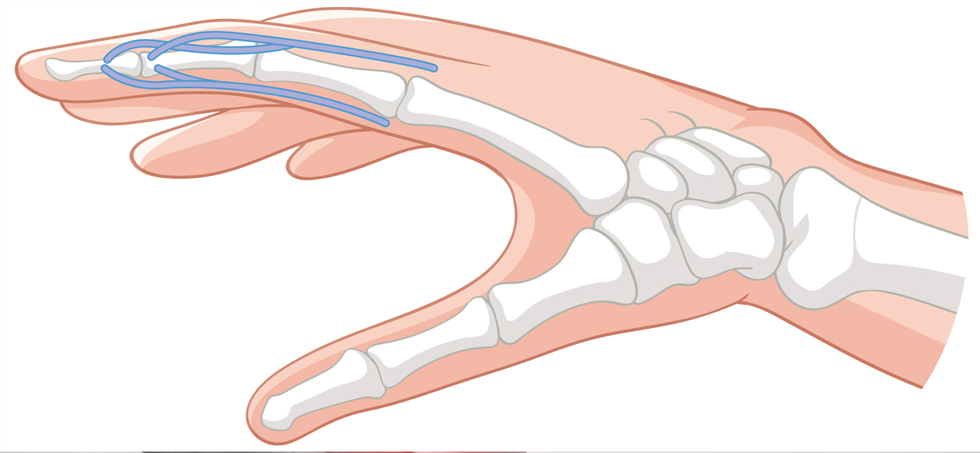
The term ’boutonnière’ is French for ‘buttonhole,’ referring to the appearance created when the central slip of the extensor tendon tears and the PIP joint protrudes through the gap between the lateral bands — resembling a button through a buttonhole.
The deformity develops when the central slip of the extensor tendon — which is responsible for straightening the PIP joint — is disrupted. This disruption causes the lateral bands to shift volarly (toward the palm), resulting in forced PIP flexion and DIP hyperextension. If left untreated for more than 3 weeks, the deformity can become fixed and significantly more difficult to correct.
Causes of Boutonniere Deformity
Boutonniere deformity may arise from a variety of traumatic, inflammatory, and congenital conditions. The most common causes include:
- Traumatic Injury: A forceful blow to the dorsal (back) surface of the middle finger joint, a laceration, crush injury, or dislocation of the PIP joint can rupture or avulse the central slip tendon. This is the most frequent cause.
- Rheumatoid Arthritis (RA): Chronic inflammation in the PIP joint leads to progressive weakening and eventual rupture of the central slip. Synovitis causes fibrous tissue buildup, bony erosion, and cartilage damage, gradually deforming the joint.
- Osteoarthritis: Degenerative changes in the finger joints can weaken the extensor mechanism and contribute to deformity over time.
- Burns: Thermal injuries to the dorsum of the finger can cause contractures and tendon damage leading to boutonniere deformity. Tension ischemia is a possible mechanism for tendon rupture in burn injuries.
- Finger Fractures: Fractures involving the middle phalanx, particularly avulsion fractures of the central slip attachment, can produce this deformity.
- Sports Injuries: Jammed fingers in contact sports such as basketball, football, and volleyball frequently involve central slip injuries that can evolve into boutonniere deformity if unrecognized.
- PIP Joint Dislocation: A volar PIP dislocation can avulse the dorsal lip of the middle phalanx, disrupting the central slip attachment.
Congenital Factors: In rare cases, congenital aplasia or hypoplasia of the finger extensors can result in boutonniere deformity from birth.
Signs and Symptoms of Boutonniere Deformity
Symptoms may develop immediately following an acute injury or may emerge gradually over 1 to 3 weeks as the lateral bands progressively migrate. Characteristic features include:
- PIP Joint Flexion Deformity: The middle joint of the affected finger is stuck in a bent position and cannot be actively straightened — the hallmark sign of boutonniere deformity.
- DIP Joint Hyperextension: The fingertip bends abnormally backward (hyperextends) due to displacement of the lateral bands and altered tendon mechanics.
- Pain and Tenderness: Pain is typically felt over the dorsal surface of the PIP joint, worsened by attempts to extend the finger.
- Swelling and Edema: Localized swelling over the middle joint of the finger, especially in the acute phase following injury.
- Loss of Grip Strength: Difficulty gripping or pinching objects due to altered finger mechanics and pain.
- Stiffness: Progressive stiffness of the PIP joint, particularly in chronic or untreated cases.
- Functional Limitation: Difficulty performing fine motor tasks such as writing, buttoning clothing, or typing.
Pathology
The pathological basis of boutonniere deformity lies in disruption of the extensor mechanism over zone III (at the level of the PIP joint). When the central slip is torn or avulsed, the lateral bands lose their dorsal stabilization and sublux volarly, passing below the axis of rotation of the PIP joint. Instead of extending the PIP joint, they now act as flexors. Simultaneously, they continue to exert their pull on the DIP joint, producing hyperextension. In rheumatoid arthritis, synovitis progressively erodes periarticular soft tissue, weakening the central slip until it ruptures under normal functional loads.
Diagnosis of Boutonniere Deformity
Prompt and accurate diagnosis is critical, as delay beyond 3 weeks significantly complicates treatment. The following diagnostic approaches are used:
Clinical Examination
A thorough history and physical examination is the primary diagnostic tool. The clinician evaluates the posture of the finger at rest, active and passive range of motion of the PIP and DIP joints, and tenderness over the central slip insertion.
Elson Test
A specific clinical test for central slip integrity. The PIP joint is held at 90° of flexion over the edge of a table and the patient is asked to extend the finger against resistance. If the DIP joint remains flexible and floppy, central slip integrity is intact. If the DIP joint becomes rigid during this maneuver, central slip disruption is confirmed. This is the most reliable bedside test for early diagnosis.
X-Ray (Radiograph)
Anteroposterior and lateral radiographs are used to identify associated avulsion fractures at the central slip insertion on the dorsal base of the middle phalanx, detect any bony fragment displacement, and assess the degree of DIP joint hyperextension on lateral views.
MRI (Magnetic Resonance Imaging)
MRI provides detailed soft tissue imaging and is useful when clinical examination is inconclusive. It can directly visualize the integrity of the central slip and surrounding structures.
Ultrasound
Dynamic ultrasound assessment can visualize the extensor tendon in real time during movement and identify central slip disruption, particularly useful in the acute clinical setting.
Treatment for Boutonniere Deformity
Treatment depends on the severity, acuity, and underlying cause of the deformity. The primary goal is to restore full active extension of the PIP joint and normal DIP joint alignment. Conservative management is preferred and most effective when initiated early.
Conservative (Non-Surgical) Treatment
- Splinting: The PIP joint is immobilized in full extension using a static splint (such as an Oval-8 or Bunnell safety-pin splint) while the DIP joint is left free to flex. Splinting is maintained continuously for 6–8 weeks for acute injuries, and up to 12 weeks for chronic cases. Allowing the finger to accidentally flex during this period can reverse all progress made.
- DIP Joint Exercises: While the PIP joint is splinted in extension, the DIP joint is actively flexed and extended throughout the day. These exercises help migrate the lateral bands dorsally back to their correct position.
- Medications: NSAIDs and analgesics for pain and inflammation management. In rheumatoid arthritis-related deformity, disease-modifying antirheumatic drugs (DMARDs) and corticosteroid injections may be used to control systemic inflammation.
- Night Splinting: After the primary immobilization period, continued night splinting may be recommended for several additional weeks to maintain PIP extension gains.
Surgical Treatment
Surgery is indicated when conservative treatment fails, when there is a severed tendon, a large displaced bony fragment, or when the deformity is caused by rheumatoid arthritis with joint destruction. Surgical options include:
- Central Slip Repair or Reconstruction: The ruptured central slip is surgically repaired using primary suture or tendon grafting. An avulsed bony fragment is reattached using a bone anchor or pin fixation.
- PIP Joint Pinning: A temporary Kirschner wire (K-wire) is inserted across the PIP joint to maintain it in full extension for 4–6 weeks during tendon healing.
- Extensor Tenotomy: In chronic boutonniere deformity, a tenotomy of the terminal extensor tendon at the DIP joint can reduce hyperextension and improve finger balance.
- Joint Replacement (Arthroplasty): For boutonniere deformity secondary to advanced rheumatoid arthritis with joint destruction, silicone PIP joint replacement may restore alignment and function.
- Joint Fusion (Arthrodesis): Fusion of the PIP joint in a functional position eliminates pain and prevents progressive deformity, at the cost of permanent loss of joint motion. Reserved for end-stage cases.
Following surgery, patients wear a splint for several weeks and undergo a structured physiotherapy program to restore mobility and strength.
Physiotherapy Treatment for Boutonniere Deformity
Physiotherapy is essential in both conservative and post-surgical management of boutonniere deformity. A personalized rehabilitation program addresses pain, inflammation, joint mobility, strength, and functional recovery.
Electrophysical Agents
- Heat Therapy: Moist heat or paraffin wax baths are applied prior to exercise sessions to warm the tissues, relax the muscles of the hand, and improve extensibility of the tendons.
- Cold Therapy / Cryotherapy: Ice packs or cold compresses are used in the acute phase to reduce swelling, pain, and local inflammation around the PIP joint.
- TENS (Transcutaneous Electrical Nerve Stimulation): Reduces pain by modulating nerve signals at the spinal cord level, allowing the patient to participate more comfortably in exercise therapy.
- Ultrasound Therapy: Therapeutic ultrasound promotes tissue healing, reduces scar tissue formation, and improves tendon extensibility in the subacute and chronic phases.
- Interferential Current Therapy (IFT): Delivers deep penetrating electrical stimulation to reduce pain, decrease swelling, and improve local circulation in the affected finger.
- Low-Level Laser Therapy (LLLT): May be used to promote cellular repair and reduce inflammation in the peritendinous tissues around the central slip.
Splinting & Orthotic Management
The physiotherapist plays a central role in fabricating and fitting custom thermoplastic splints to maintain the PIP joint in full extension. Splinting protocols are progressively modified as healing advances — transitioning from static to dynamic splints that allow controlled range of motion while continuing to protect the healing tendon.
Exercise Therapy
- DIP Flexion Exercises (During Splinting): With the PIP joint held in extension by the splint, the patient actively flexes and extends the DIP joint repeatedly throughout the day to maintain DIP mobility and promote dorsal migration of the lateral bands.
- PIP Extension Exercises: After the immobilization period, gentle active-assisted and active PIP extension exercises are introduced to restore extensor tendon function and joint range of motion.
- Tendon Gliding Exercises: A sequence of specific hand positions (straight, hook fist, full fist, tabletop) performed repetitively to promote independent gliding of the flexor and extensor tendons and prevent adhesion formation.
- Strengthening Exercises: Progressive resistance exercises using putty, grip strengtheners, and squeeze balls to restore grip strength, pinch strength, and intrinsic muscle function.
- Stretching Exercises: Gentle passive stretching of the PIP joint into extension and the DIP joint into flexion to restore the balance between flexor and extensor forces.
Manual Therapy
- Joint Mobilization: Skilled passive mobilization techniques applied to the PIP and DIP joints to improve joint capsule extensibility and restore physiological range of motion.
- Soft Tissue Massage: Massage and myofascial release techniques applied to the intrinsic muscles of the hand, thenar and hypothenar eminences, and the dorsal tendon structures to reduce tension and improve blood flow.
- Scar Management: In post-surgical or post-laceration cases, scar massage, silicone gel sheeting, and desensitization techniques are employed to minimize adhesion formation and improve scar pliability.
Functional Rehabilitation
As recovery progresses, functional retraining focuses on restoring the ability to perform activities of daily living (ADLs). Task-specific activities such as pinching, grasping, writing, and typing are progressively reintroduced to retrain coordinated finger function.
Occupational Therapy Integration
Occupational therapists work alongside physiotherapists to address ADL performance, adaptive equipment, workplace modifications, and vocational rehabilitation for patients whose daily activities or occupation have been impacted by the deformity.
Patient Education
Education is an integral part of recovery from boutonniere deformity. Patients are counseled on the following:
- Understanding the Condition: Clear explanation of the anatomy of the extensor mechanism, the mechanism of injury, and why consistent splinting and exercises are essential to recovery.
- Importance of Early Treatment: Patients are informed that delaying treatment beyond 3 weeks significantly worsens outcomes and may make the deformity permanent or require more complex surgical intervention.
- Splint Compliance: Emphasis on wearing the splint continuously as prescribed — removing it to bathe or clean the splint — and never allowing the finger to flex at the PIP joint during the immobilization period, as even a single accidental bend can restart the healing clock.
- Home Exercise Program: Instruction in a personalized set of daily exercises including DIP flexion, tendon gliding, and progressive strengthening to be performed independently between clinic sessions.
- Activity Modification: Guidance on avoiding activities that risk re-injury to the finger during the healing phase. Return to sports is permitted only with appropriate protective splinting or buddy taping.
- Recognizing Warning Signs: Patients are educated on signs of deterioration — worsening deformity, increased pain, or numbness — that warrant prompt reassessment by their healthcare provider.
- Long-term Prognosis: Realistic discussion of recovery timelines (3–6 months for full recovery), the potential for residual stiffness, and the importance of ongoing hand exercises after formal rehabilitation ends.
Related Conditions
Boutonniere deformity shares clinical overlap with and may be associated with the following conditions:
- Rheumatoid Arthritis: A major systemic cause of boutonniere deformity through progressive synovitis and central slip erosion.
- Swan Neck Deformity: The inverse finger deformity — PIP hyperextension and DIP flexion — also commonly seen in rheumatoid arthritis.
- Mallet Finger: Disruption of the terminal extensor tendon causing DIP flexion deformity; a related extensor tendon injury.
- Trigger Finger (Stenosing Tenosynovitis): Inflammation of the flexor tendon sheath causing locking or catching of the finger in flexion.
- Dupuytren’s Contracture: Progressive fibrosis of the palmar fascia causing fixed flexion deformity of the fingers.