WHAT IS AN ANKLE BONE SPUR?
An Ankle Bone Spur, clinically identified as an Osteophyte, represents a common yet often misunderstood orthopedic condition. These are essentially extra growths of bone tissue that develop on the edges of the ankle joint. While the word “spur” often evokes the image of a sharp, needle-like projection, most bone spurs are actually smooth, rounded mounds of calcium and bone that form over long periods.
At Physiotherapy Expert, we encounter patients from all walks of life—from professional athletes suffering from “Footballer’s Ankle” to older adults experiencing the natural degenerative changes of “Wear and Tear.” Our philosophy is built on the belief that while you cannot “dissolve” a bone spur through exercise, you can absolutely retrain the joint to move in a way that makes the spur asymptomatic. This comprehensive guide explores the biological mechanisms of bone growth, the diagnostic process, and the advanced physiotherapy interventions we use to restore mobility.
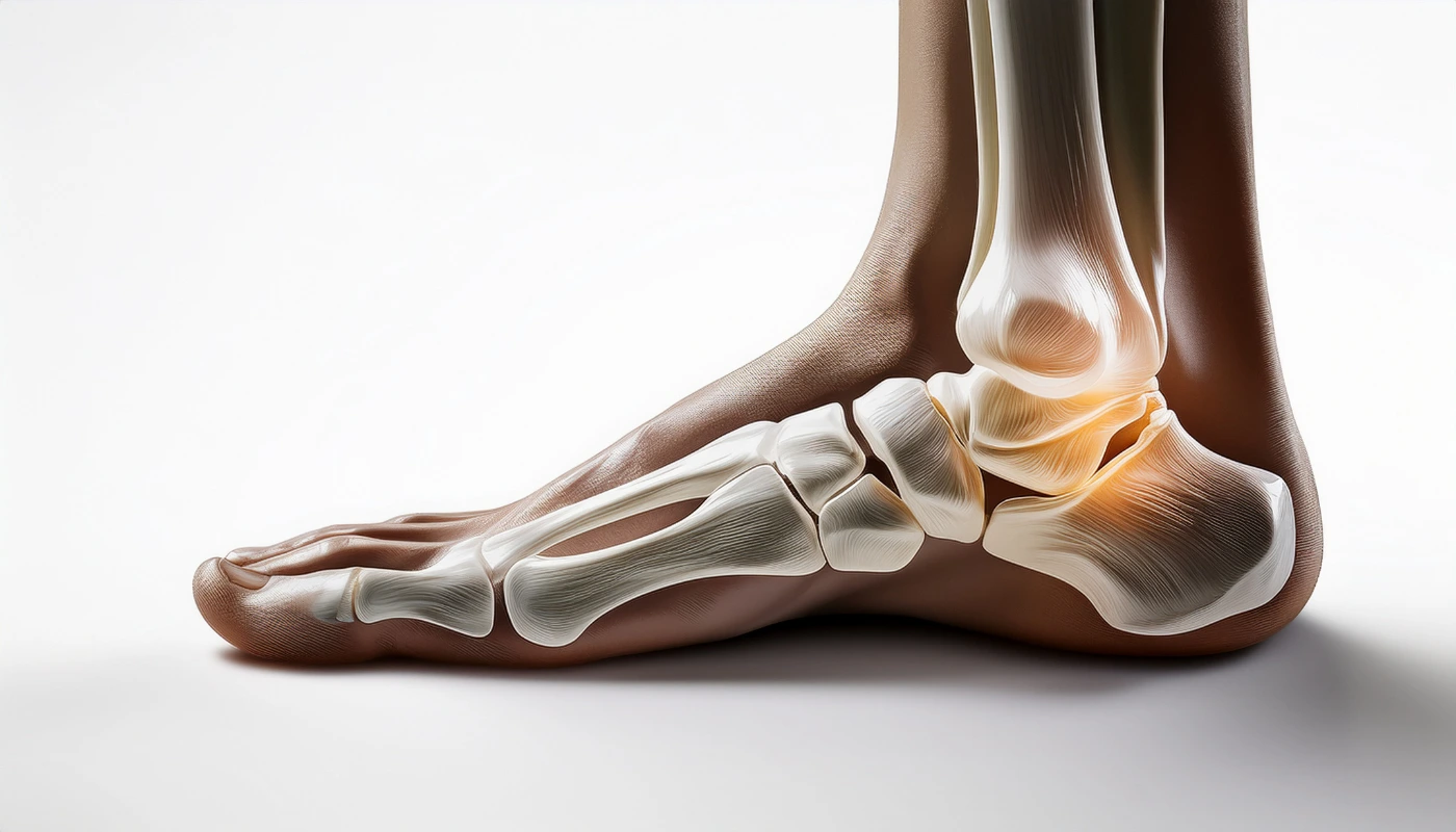
A bone spur is the body’s adaptive, biological response to persistent stress, friction, or pressure. In the context of the ankle, these growths typically manifest where two or more bones meet (the joint interface). The ankle joint, or the talocrural joint, is a complex hinge involving the Tibia (shinbone), the Fibula (outer leg bone), and the Talus (the bone that connects the leg to the foot).
When the protective hyaline cartilage that caps these bones begins to thin or fray—whether due to age, injury, or repetitive impact—the body senses an increase in joint instability. To compensate, the periosteum (the membrane covering the bone) triggers the production of new bone cells to spread the weight over a larger surface area. These resulting “bumps” are osteophytes.
When these spurs grow on the front (anterior) part of the ankle, they can cause Anterior Ankle Impingement. This is a mechanical “pinching” sensation that occurs when you try to pull your toes toward your shin, a movement known as dorsiflexion.
COMMON CAUSES OF ABNORMAL BONE GROWTH
Bone spurs are rarely the primary problem; they are usually a symptom of an underlying mechanical issue. Understanding the “why” behind the growth is the first step in the Physio Expert assessment.
1. Ankle Osteoarthritis
The most frequent driver of osteophyte formation is osteoarthritis. As the joint space narrows and the cartilage wears down, the bones begin to grate against one another. The inflammation from this friction signals the bone-building cells (osteoblasts) to create spurs to “stiffen” the joint and protect it from further movement that might cause damage.
2. Repetitive Trauma (Footballer’s Ankle)
This condition is prevalent in athletes who perform repetitive, forceful kicking or extreme bending of the ankle. In soccer players, the constant tension on the ligaments that attach to the bone causes the bone to pull outward, stimulating growth. Similarly, dancers who perform “en pointe” or deep pliés put immense pressure on the front and back of the ankle, leading to impingement spurs.
3. Chronic Ankle Instability
If you have a history of multiple ankle sprains, your ligaments may have become “lax” or loose. This creates micro-motions within the joint that shouldn’t be there. The body tries to stabilize this “wobble” by growing bone spurs around the edges of the joint to act as natural stoppers.
4. Natural Aging and Genetics
For some individuals, bone spurs are simply a byproduct of the aging process. As we age, our ligaments lose elasticity and our bone metabolism changes. Additionally, some people are genetically predisposed to forming bone more readily in response to minor irritations.
SYMPTOMS: RECOGNIZING THE SIGNS OF IMPINGEMENT
Many people have bone spurs in their ankles and never know it. A spur only becomes a “condition” when it begins to irritate the surrounding soft tissues (tendons, nerves, or the joint capsule).
- Mechanical Blockage: One of the most telling signs is the feeling that your ankle “hits a wall” when you try to squat, climb stairs, or lunge.
- Aching Pain: A dull, persistent ache deep within the joint that worsens after long periods of standing or walking.
- Sharp, Pinching Pain: Specifically at the front of the ankle joint line during activities that require the shin to move over the foot.
- Visible or Palpable Bumps: You may feel a hard, immovable knot under the skin, particularly on the top of the foot where it meets the leg.
- Swelling: Chronic irritation from the spur rubbing against the synovium (joint lining) can cause “synovitis,” leading to a puffy, swollen appearance around the ankle “knobs” (malleoli).
DIAGNOSIS: BEYOND THE SURFACE
At Physiotherapy Expert, we believe a diagnosis is a roadmap for treatment. We use a combination of physical testing and advanced imaging to build your profile.
- Clinical Impingement Test: We perform a “forced dorsiflexion” test. If this reproduces your specific sharp pain, it is a strong indicator of anterior impingement.
- Weight-Bearing X-Rays: An X-ray is the most effective tool for visualizing bone. We specifically look at “lateral” (side-view) X-rays to see the height and shape of the spurs on the talus and tibia.
- MRI (Magnetic Resonance Imaging): While X-rays show the bone, an MRI shows the “cost” of the spur. It allows us to see if the spur is fraying the nearby tendons or if there is “bone marrow edema” (bruising inside the bone) caused by the spurs knocking together.
PHYSIOTHERAPY TREATMENT: THE PHYSIO EXPERT PROTOCOL
Our approach to ankle bone spurs is focused on Joint Decompression. Since we cannot remove the bone, we focus on creating more “space” within the joint so the spur has room to move without pinching.
Phase 1: The De-Inflammation Stage
When you first visit us, your ankle is likely in a state of high irritation.
- Manual Therapy (Joint Distraction): Our therapists use hands-on techniques to gently pull the foot away from the leg. This “un-weights” the joint and can provide immediate, temporary relief from the pinching sensation.
- Cryotherapy: We use specialized ice protocols to calm the synovitis (inflammation of the joint lining) caused by the spur.
- Activity Modification: We teach you how to maintain your fitness (using swimming or cycling) while avoiding the specific “end-range” movements that trigger the spur.
Phase 2: Restoring Mechanical Harmony
- Posterior Chain Mobility: If your calf muscles are tight, they pull the ankle into a position that makes impingement more likely. We use deep tissue release and eccentric stretching to lengthen the calves.
- Taping for Decompression: We use Kinesiology tape to create a “lift” in the skin and soft tissues, which can subtly change the pressure markers within the ankle joint.
- Heel Lifts and Footwear Advice: Sometimes, a simple 5mm heel lift inside your shoe can tilt the ankle just enough to prevent the spur from making contact with the tibia during walking.
Phase 3: Stabilization and Load Management
- Proprioception Training: We use wobble boards and BOSU balls to sharpen your “joint position sense.” A stable ankle is less likely to experience the micro-shifts that cause the spur to pinch.
- Strengthening the Intrinsic Muscles: We focus on the tiny muscles inside the foot that support the arch. A strong arch provides a better mechanical base for the ankle joint.
- Load Progression: We gradually reintroduce impact activities (like running or jumping) using a “symptom-guided” approach to ensure the bone growth isn’t being re-irritated.
THE ROLE OF TECHNOLOGY IN BONE SPUR RECOVERY
At Physiotherapy Expert, we supplement traditional manual therapy with modern medical technology:
- Laser Therapy: Cold laser treatment is excellent for reducing the chronic, deep-seated pain of bone-on-bone irritation without the side effects of anti-inflammatory drugs.
- Extracorporeal Shockwave Therapy (ESWT): While primarily used for tendons, shockwave can be effective in desensitizing the nerves around a bone spur, significantly reducing pain levels.
- Ultrasound Therapy: We use therapeutic ultrasound to provide deep heating to the joint capsule, making our manual stretching techniques more effective.
SURGICAL INTERVENTION: WHEN IS IT NECESSARY?
Most patients with ankle bone spurs find relief through physiotherapy. However, surgery (Ankle Debridement) may be considered if:
- The spur is physically preventing you from performing your job or sport after 6 months of dedicated rehab.
- The spur has broken off (forming a “loose body”) and is locking the joint.
- There is significant nerve entrapment.
Post-surgical physiotherapy is arguably more important than the surgery itself. Without proper rehab, scar tissue can form in the space where the bone was removed, leading to a return of the impingement symptoms.
FREQUENTLY ASKED QUESTIONS
1. Can I “rub out” a bone spur with massage?
2. Why does my ankle hurt more in the morning?
3. Are bone spurs the same as heel spurs?
4. Can certain shoes cause bone spurs?
PREVENTING FUTURE BONE GROWTH
While you may already have a spur, you can prevent it from getting larger or prevent new ones from forming:
- Maintain a Healthy Weight: Every extra pound increases the force across the small ankle joint by four-fold during walking.
- Wear Supportive Footwear: Ensure your shoes have adequate cushioning and are replaced once the midsoles compress.
- Never “Push Through” Pinching Pain: If you feel a sharp pinch in your ankle during exercise, stop and adjust your position. “No pain, no gain” does not apply to mechanical impingement.