WHAT IS RETROLISTHESIS?
Retrolisthesis is a clinical condition of the spine characterized by the posterior or “backward” displacement of one vertebral body in relation to the vertebra immediately below it. While many patients are familiar with its more common counterpart, Spondylolisthesis (the forward slippage of a vertebra), Retrolisthesis presents its own unique set of mechanical and neurological challenges. This structural shift is rarely an isolated event; it is often the hallmark of localized spinal instability, ligamentous laxity, or advanced degenerative changes within the intervertebral discs.
At Physiotherapy Expert, we approach Retrolisthesis through the lens of Segmental Spinal Stabilization. We believe that while a structural “slip” is captured on an X-ray, the true pathology lies in the failure of the deep stabilizing muscles to maintain the integrity of the spinal column. Our mission is to transform a vulnerable, unstable spine into a resilient, supported structure through evidence-based manual therapy, precise corrective exercise, and advanced medical technology.
The human spine is a marvel of biological engineering, designed to provide a rigid protective housing for the spinal cord while allowing for multidirectional flexibility. In a healthy spine, the anterior (front) and posterior (back) walls of the vertebral bodies align perfectly, forming a smooth, continuous column. Retrolisthesis occurs when this alignment is breached.
When a vertebra moves backward, even by a few millimeters, it alters the dimensions of the spinal canal and the neural foramina (the openings where nerves exit the spine). This displacement can lead to “mechanical” pain from the strained ligaments and “neurological” pain from the compression of nerve roots. At Physio Expert, we categorize Retrolisthesis based on the region of the spine it affects:
- Lumbar Retrolisthesis: Most common in the L4-L5 or L5-S1 segments due to the immense weight-bearing demands of the lower back.
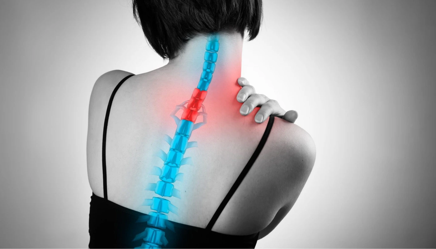
- Cervical Retrolisthesis: Often found in the neck (C3-C5), frequently resulting from whiplash injuries or chronic “tech-neck” posture.
Thoracic Retrolisthesis: Rare, due to the stabilizing effect of the rib cage, but highly significant when present.
UNDERSTANDING THE GRADING SYSTEM
To provide a precise prognosis and build a safe rehabilitation roadmap, we utilize a grading system based on the percentage of the posterior slip relative to the width of the vertebral body:
- Grade 1: Up to 25% displacement. This is the most common presentation and responds exceptionally well to conservative physiotherapy.
- Grade 2: 26% to 50% displacement. This involves significant stretching of the anterior longitudinal ligament and requires intensive, long-term stabilization.
- Grade 3: 51% to 75% displacement. At this stage, spinal stability is severely compromised, often resulting in marked neurological deficits.
- Grade 4: 76% to 100% displacement. This is a critical stage (Complete Retrolisthesis) that usually necessitates surgical intervention alongside post-operative rehabilitation.
At Physio Expert, our first step is a thorough review of your imaging (X-ray, CT, or MRI) to determine your grade and ensure that our exercise load does not exceed the structural tolerance of your spine.
COMMON CAUSES OF VERTEBRAL DISPLACEMENT
Vertebrae do not shift backward without a breakdown in the spine’s primary or secondary stabilizers. The most common causes identified in our clinic include:
1. Degenerative Disc Disease (DDD)
The intervertebral disc acts as a hydraulic shock absorber. As we age, these discs lose water content (desiccation) and height. As the disc space narrows, the ligaments surrounding the joint become “slack,” allowing the vertebra to slide backward under the influence of gravity and muscle tension.
2. Traumatic Injury
A sudden, high-velocity force—such as a motor vehicle accident or a significant fall onto the tailbone—can cause an acute shear force that overcomes the strength of the spinal ligaments, leading to an immediate displacement.
3. Facet Joint Arthritis
The facet joints at the back of the spine provide the “tracks” for movement. When these joints develop arthritis (hypertrophy), they can no longer effectively block the backward sliding of the vertebra.
4. Core Muscle Atrophy
The deep stabilizers—specifically the Multifidus and Transverse Abdominis—act as an internal corset. When these muscles are weak due to a sedentary lifestyle or chronic pain, the “active” stability of the spine is lost, leaving the bones vulnerable to shifting.
SYMPTOMS: RECOGNIZING THE SIGNS OF INSTABILITY
Retrolisthesis symptoms can be deceptive, often mimicking a simple “pulled muscle” or a herniated disc. Key indicators include:
- Localized Deep Ache: A persistent, “boring” pain directly over the site of the slip.
- Neurogenic Claudication: Pain, numbness, or heaviness in the legs that worsens with walking or standing and improves when sitting or leaning forward.
- Radiculopathy (Sciatica): Sharp, shooting pains that travel into the buttocks, thighs, or calves if the displaced bone is pinching a nerve root.
- Reduced Range of Motion: A noticeable “catch” or sharp pain when trying to arch the back or twist.
- Muscle Spasms: The large muscles of the back (Erector Spinae) often go into a protective “guarding” state to try and hold the spine still.
DIAGNOSIS: THE CLINICAL BLUEPRINT
At Physiotherapy Expert, we don’t just look at the bone; we look at the person. Our diagnostic process involves:
- Static Palpation: We feel for a “step-off” deformity—a palpable dip in the spine where the vertebra has shifted.
- Neurological Screening: Testing your reflexes, dermatomes (sensation), and myotomes (strength) to determine if the Retrolisthesis is impacting the spinal cord or nerves.
- Functional Movement Assessment: Observing how your spine behaves during a squat, a bend, or a lift to identify “instability catches.”
- Radiology Review: We analyze weight-bearing lateral X-rays, which are the gold standard for measuring the exact degree of posterior slippage.
PHYSIOTHERAPY TREATMENT AT PHYSIO EXPERT: THE 12-WEEK STABILIZATION PROTOCOL
Our rehabilitation program for Retrolisthesis is scientifically designed to progress from pain relief to total spinal resilience.
Phase 1: Pain Modulation & Neural Protection (Weeks 1–4)
The immediate priority is to calm the nervous system and reduce the “inflammatory soup” around the displaced segment.
- Postural Education: We teach “Neutral Spine” mechanics. For Retrolisthesis, we often advise avoiding excessive “extension” (arching the back), which can further jam the displaced bone into the nerve space.
- Manual Therapy: Using gentle, oscillatory mobilizations to reduce muscle guarding without putting shearing force on the unstable segment.
- Cryotherapy & Laser Therapy: Utilizing medical-grade cold therapy and laser to reduce deep-seated inflammation in the ligaments and facet joints.
- Deep Core Activation: Teaching the “abdominal hollow” and “bracing” techniques to provide immediate internal support.
Phase 2: Segmental Stabilization (Weeks 4–8)
Once the acute pain is managed, we focus on the “active” stabilizers of the spine.
- Multifidus Training: The Multifidus is the most important muscle for stopping vertebral slippage. We use targeted exercises to re-engage these tiny muscles.
- Dynamic Stability: Exercises like the “Dead Bug” and “Bird-Dog” challenge the spine to stay still while the arms and legs move.
- Pelvic Tilt Correction: Many Retrolisthesis patients have an exaggerated pelvic tilt. We balance the hip flexors and glutes to level the pelvis and reduce the backward “pull” on the lumbar vertebrae.
Phase 3: Functional Load & Durability (Weeks 8–12)
The final stage prepares you for the demands of real life—lifting, twisting, and sports.
- Proprioceptive Training: Using unstable surfaces to sharpen the brain’s ability to keep the spine aligned during sudden movements.
- Progressive Loading: Introducing weighted carries and deadlift variations (with strict form) to build the “armour” of muscle around the spine.
- Ergonomic Integration: Tailoring your workstation and daily habits to ensure your new spinal stability is maintained for years to come.
THE PHYSIO EXPERT ADVANTAGE: TECHNOLOGY IN SPINAL REHAB
- Neuromuscular Electrical Stimulation (NMES): We use NMES to “force” the deep stabilizers to contract in cases where the patient’s brain-to-muscle connection has been severed by chronic pain.
- Kinesiology Taping: Providing a 24/7 “tactile reminder” to the brain to keep the spine in a neutral, safe position.
- Traction (Intermittent): In cases of severe nerve entrapment, manual traction can provide a “window of relief” by temporarily increasing the space in the neural foramina.
POTENTIAL COMPLICATIONS AND VIGILANCE
Retrolisthesis, if left unmanaged, can lead to secondary conditions:
- Spinal Stenosis: The backward slip reduces the diameter of the spinal canal, leading to chronic leg heaviness and walking difficulties.
- Foraminal Narrowing: The “exit doors” for the nerves become cramped, leading to permanent nerve irritability.
- Spondylosis: Accelerated “wear and tear” on the adjacent vertebrae as they work harder to compensate for the unstable segment.
FREQUENTLY ASKED QUESTIONS
1. Can a bone “slip back” into place with physiotherapy?
2. Is surgery the only permanent fix?
3. What exercises should I avoid?
4. How long does it take to feel better?
RELATED CONDITIONS
- Herniated Disc: Often occurs at the same level as a Retrolisthesis due to the abnormal shear forces on the disc.
- Spondylolysis: A stress fracture in the “pars” of the vertebra that can lead to slippage.
- Sciatica: Nerve pain originating from the lumbar spine that frequently accompanies vertebral displacement.