
WHAT IS LUMBAR SPONDYLOSIS?
Lumbar Spondylosis is a broad medical term used to describe the age-related, progressive degeneration of the lumbar spine (lower back). It is a form of spinal osteoarthritis that specifically targets the intervertebral discs, vertebral bodies, and the associated facet joints of the five lumbar vertebrae ($L1$ to $L5$). As we age, the structural integrity of the spine naturally declines, leading to bone spurs, disc thinning, and ligamentous thickening.
At Physio Expert, we view Lumbar Spondylosis not merely as an “inevitable part of aging,” but as a manageable mechanical condition. While the structural changes are often irreversible, the functional limitations—such as pain, stiffness, and reduced mobility—can be significantly improved through targeted, evidence-based physiotherapy. Our goal is to stabilize the spine, decompress neural structures, and restore the patient’s quality of life.
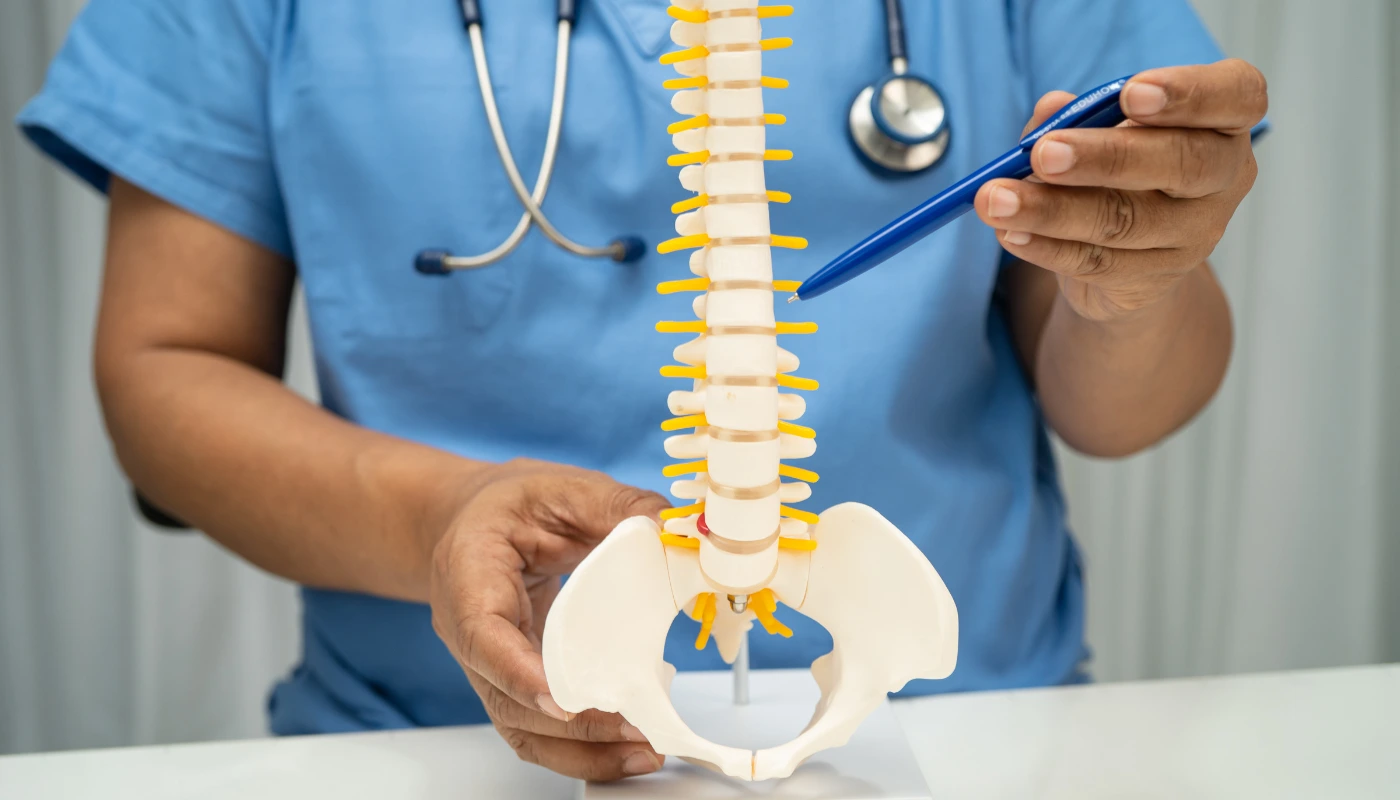
The Anatomy of the Lumbar Spine
The lumbar spine is designed to bear the majority of the body’s weight and provide a wide range of motion. Each segment consists of a vertebral body, a shock-absorbing intervertebral disc, and two facet joints at the back that guide movement. In Lumbar Spondylosis, these components begin to break down in a “cascade of degeneration,” where a problem in one part of the segment inevitably leads to stress in the others.
WHAT ARE THE CAUSES OF LUMBAR SPONDYLOSIS?
Lumbar Spondylosis is a multifactorial condition. While aging is the primary driver, several other factors accelerate the wear and tear of the spine.
1. Age-Related Wear and Tear
By the age of 60, over 80% of individuals show some radiographic evidence of spondylosis. Over decades, the constant mechanical loading of the spine leads to micro-trauma and structural fatigue.
2. Genetic Predisposition
Heredity plays a significant role in the quality of your collagen and bone density. If your parents suffered from early-onset back pain or “slipped discs,” you may be genetically predisposed to faster spinal degeneration.
3. Occupational and Mechanical Stress
Jobs that require repetitive heavy lifting, prolonged sitting, or frequent twisting and bending place abnormal shear forces on the lumbar discs. Professional drivers, construction workers, and office employees with poor ergonomics are at high risk.
4. Previous Spinal Trauma
A history of car accidents, sports injuries, or falls can create localized instability in the spine. Even if the injury healed years ago, the altered biomechanics often lead to accelerated spondylosis in the affected segments.
5. Obesity and Lifestyle
Excess body weight increases the vertical load on the lumbar vertebrae. Furthermore, a sedentary lifestyle leads to “disuse atrophy” of the core muscles, leaving the bony spine to support the entire weight of the torso without muscular assistance.
WHAT ARE THE SYMPTOMS OF LUMBAR SPONDYLOSIS?
The symptoms of Lumbar Spondylosis vary based on the severity of the degeneration and whether the nerves are involved.
- Chronic Lower Back Pain: Persistent, dull aching in the lumbar region that often worsens after prolonged activity or sitting.
- Morning Stiffness: A “locked” feeling in the back upon waking that gradually improves with movement and heat.
- Neurogenic Claudication: Pain, numbness, or heaviness in the legs that occurs while walking or standing and is relieved by sitting or leaning forward (the “shopping cart” sign).
- Radiculopathy (Sciatica): Sharp, shooting pain that radiates down the buttocks into the legs, caused by bone spurs pressing on spinal nerves.
- Muscle Weakness: Difficulty in performing tasks like rising from a chair or walking on heels/toes due to nerve root compression.
- Crepitus: A grinding or popping sensation during spinal movement, signifying bone-on-bone friction in the facet joints.
- Disc Bulging Symptoms: Sharp pain during coughing, sneezing, or straining, which increases intradiscal pressure.
PATHOLOGY: THE DEGENERATIVE CASCADE
The pathology of Lumbar Spondylosis follows a predictable clinical path often referred to as Kirkaldy-Willis’s Three-Phase Model.
Phase 1: Dysfunction
Degeneration begins with the dehydration of the Nucleus Pulposus (the jelly-like center of the disc). As it loses water, the disc height decreases, causing the surrounding Annulus Fibrosus (the outer ring) to develop small tears.
Phase 2: Instability
As disc height continues to drop, the ligaments (Ligamentum Flavum) become lax. The facet joints, which are not designed to bear weight, begin to take on the load. This leads to inflammation, cartilage loss, and hypermobility of the spinal segment.
Phase 3: Stabilization (The Formation of Osteophytes)
In a desperate attempt to stabilize the wobbly segment, the body grows extra bone. These are known as Osteophytes or bone spurs. While they provide some stability, they often grow into the spinal canal (stenosis) or the exit holes for nerves (foraminal stenosis), leading to chronic nerve pain. At Physio Expert, our pathological goal is to bypass this instability by building a “muscular corset” to support the spine.
DIAGNOSIS OF LUMBAR SPONDYLOSIS
Accurate diagnosis is vital to differentiate spondylosis from other conditions like kidney stones or vascular issues.
1. Comprehensive Physical Examination
We assess the range of motion, perform provocative tests (like the Straight Leg Raise), and evaluate neurological reflexes to pinpoint the affected spinal level.
2. X-Ray Radiography
X-rays are the first line of defense. They clearly show narrowed disc spaces, bone spurs (osteophytes), and facet joint thickening.
3. MRI (Magnetic Resonance Imaging)
MRI is the “gold standard” for visualizing soft tissues. It allows us to see the exact degree of disc bulging, the health of the spinal cord, and the extent of nerve root “pinching.”
4. CT Scan & SPECT
CT scans provide a detailed cross-sectional view of the bone. SPECT (Single-photon emission computed tomography) is a specialized nuclear scan used to identify “active” areas of inflammation in the facet joints, helping us target treatment more effectively.
TREATMENT OF LUMBAR SPONDYLOSIS
Medical Management
- NSAIDs: To manage chronic inflammation.
- Muscle Relaxants: For acute spasms that often accompany spondylosis flare-ups.
- Epidural Steroid Injections: To deliver powerful anti-inflammatories directly to the irritated nerve roots.(Note: Medications should only be used as a bridge to allow for active physiotherapy.)
Surgical Management
Surgery is considered a last resort when there is progressive neurological loss (e.g., foot drop) or bowel/bladder dysfunction.
- Laminectomy: Removing the back of the vertebra to relieve pressure.
- Discectomy: Removing the bulging part of the disc.
- Spinal Fusion: Welded vertebrae together to eliminate painful movement in an unstable segment.
PHYSIOTHERAPY TREATMENT AT PHYSIO EXPERT
Our rehabilitation strategy is built on a four-phase model designed to take you from acute pain to functional independence.
Phase 1: Pain Alleviation & Offloading (Weeks 1–2)
The priority is to break the pain-spasm cycle.
- Cryotherapy & Thermotherapy: We use ice to reduce acute inflammation and heat to relax guarded muscles.
- TENS & Interferential Therapy (IFT): Advanced electrical stimulation to block pain signals and reduce the need for oral medication.
- Kinesio-Taping: Specialized taping techniques to support the lower back muscles and provide sensory feedback for better posture.
- Mechanical Traction: Using a traction table to gently “stretch” the lumbar spine, opening the intervertebral spaces and reducing pressure on the discs.
Phase 2: Mobility & Myofascial Release (Weeks 3–5)
Once pain is managed, we address the “tightness” that keeps the spine in a compressed state.
- Manual Therapy: Spinal mobilization (Grade I–IV) to improve joint lubrication and break down adhesions.
- Soft Tissue Massage: Releasing the Erector Spinae and Quadratus Lumborum muscles that are often chronically tight.
- Stretching Exercises: Focused on the Hamstrings, Hip Flexors, and Piriformis. Tightness in these areas causes the pelvis to tilt, which increases the stress on the lumbar vertebrae.
Phase 3: Core Stabilization & Strengthening (Weeks 6–9)
We focus on the “Deep Core”—the muscles that act as your internal back brace.
- McKenzie Exercises: Specific extension-based movements that help “centralize” disc-related pain and restore the natural lumbar curve (lordosis).
- The “Big 3” Core Routine: Exercises like the Bird-Dog, Side Plank, and Modified Curl-up to build endurance in the spinal stabilizers.
- VMO & Gluteal Activation: Strengthening the hips to ensure they take the load during daily activities, sparing the lower back.
- Neuromuscular Re-education: Using biofeedback to ensure the patient is engaging the correct muscles during movement.
Phase 4: Functional Loading & Ergonomic Integration (Weeks 10–12+)
- Gait Training: Correcting walking patterns to reduce the impact on the lumbar joints.
- Lifting Mechanics: Re-training the “hip hinge” to ensure the patient lifts using the legs rather than the spine.
- Proprioception & Balance: Using wobble boards to improve the body’s subconscious ability to stabilize the spine during unexpected movements.
- Long-Term HEP (Home Exercise Program): A customized, lifelong routine to prevent future flare-ups.
THE PHYSIO EXPERT ADVANTAGE: ADVANCED MODALITIES
We utilize state-of-the-art technology to accelerate the healing of degenerative tissues:
- Laser Therapy: High-intensity laser to stimulate cellular repair in the facet joints.
- PEMF (Pulsed Electromagnetic Field): To promote bone and cartilage health.
- Dry Needling: To deactivate deep-seated trigger points in the paraspinal muscles.
PATIENT EDUCATION: PROTECTING YOUR SPINE FOR LIFE
At Physio Expert, we believe an educated patient is a resilient patient.
1. Posture and Ergonomics
We teach you the “Dynamic Posture.” The spine is not meant to be static. We provide guidance on workstation setup, lumbar rolls for chairs, and the importance of taking “micro-breaks” every 30 minutes.
2. Weight Management and Nutrition
Excess abdominal weight acts as a “lever” that pulls the spine forward. We provide basic nutritional advice focused on anti-inflammatory foods (Omega-3s, Turmeric) and Vitamin D for bone health.
3. Sleep Hygiene
We recommend sleeping on your side with a pillow between your knees or on your back with a pillow under your knees. This keeps the lumbar spine in a “neutral” position and prevents morning stiffness.
4. Smoking Cessation
Smoking restricts blood flow to the spinal discs, accelerating the dehydration of the nucleus pulposus. Quitting is essential for anyone suffering from Lumbar Spondylosis.
RELATED CONDITIONS
- Lumbar Stenosis: Narrowing of the spinal canal often caused by spondylosis.
- Spondylolisthesis: The slipping of one vertebra over another.
- Degenerative Disc Disease (DDD): The primary component of spondylosis.
- Sciatica: Nerve pain resulting from spinal degeneration.